Chapter 16
Congenital Lung Disease
Learning Objectives
- Recognize venolobar syndrome on a frontal chest radiograph, chest computed tomography (CT), and chest magnetic resonance image and explain the etiology of the retrosternal band of opacity seen on the lateral radiograph.
- Name the components of pulmonary venolobar syndrome.
- Recognize a mass in the posterior segment of a lower lobe on a chest radiograph and CT and suggest the possible diagnosis of pulmonary sequestration.
- Describe the differences between intralobar and extralobar sequestration.
- Recognize bronchial atresia on a chest radiograph and CT and name the most common lobes in which it occurs.
- Recognize a cystic mass in the mediastinum and suggest the possible diagnosis of a bronchogenic cyst.
The most common major anomalies of pulmonary development (Table 16-1) span a continuum of maldevelopment involving the pulmonary parenchyma, the pulmonary vessels, or a combination of both (1).
At one end of the spectrum, congenital lobar emphysema represents
abnormal lung supplied by normal vessels, and at the other end,
pulmonary arteriovenous malformation (AVM) consists of abnormal vessels
within normal lung parenchyma. Some patients with congenital lung
anomalies have mixed features, making exact categorization of the
anomaly difficult.
Congenital Lobar Emphysema
Congenital lobar emphysema (CLE) is a disorder affecting
neonates and young infants and is usually associated with acute or
subacute respiratory distress. It may occasionally present as an
incidental finding in adults. Various bronchial and alveolar
abnormalities can cause this disorder, and in some cases the cause is
unknown. The most commonly detected abnormality is absence or
hypoplasia of cartilage rings of major and branch bronchi, with
resultant bronchial collapse during exhalation. This results in
inhalational air entry but collapse of the narrow bronchial lumen
during exhalation. The bronchial obstruction leads to progressive
hyperinflation and air trapping (Fig. 16-1),
usually involving only one pulmonary lobe. The left upper lobe is most
commonly involved, followed by the right middle and right upper lobes.
CLE has two forms: hypoalveolar (fewer than expected number of alveoli)
and polyalveolar (greater than expected number of alveoli). The
pulmonary vasculature, although frequently attenuated, is usually
normal in structure and distribution. Common chest radiographic
findings include a hyperlucent lobe, compressive atelectasis of
adjacent parenchyma, and contralateral mediastinal shift (1).
In some cases, a subtle hyperlucent lobe may be all that is seen.
Computed tomography (CT) best characterizes this abnormality and
typically shows a hyperlucent, hyperexpanded lobe;
attenuated but intact pattern of vascularity; compression of the adjacent lung; and contralateral mediastinal shift.
P.265
attenuated but intact pattern of vascularity; compression of the adjacent lung; and contralateral mediastinal shift.
TABLE 16-1 MAJOR ANOMALIES OF PULMONARY DEVELOPMENT | |
|---|---|
|
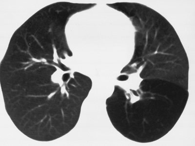 |
FIGURE 16-1. Congenital lobar emphysema.
CT of a 30-year-old asymptomatic woman shows abnormal lucency and
diminutive vasculature in the superior segment of the left lower lobe.
There was no evidence of endobronchial lesion on CT or bronchoscopy,
and the appearance of the left lower lobe was unchanged for 3 years on
follow-up CT scans. |
Bronchogenic Cyst
Bronchogenic cysts result from abnormal growth of the
lung bud and can be either mediastinal or intrapulmonary. Both types
are lined with ciliated columnar epithelium and contain serous or
mucous material. No clear-cut predilection for an intrapulmonary or a
mediastinal location has been demonstrated. An intrapulmonary
bronchogenic cyst often communicates with the bronchial tree, which can
result in air–fluid levels and recurrent infections that may damage the
cyst wall. Chest radiographs show a nonspecific oval or round,
well-circumscribed mass, often as an incidental finding. The most
common mediastinal location is subcarinal (Figs. 6-29, 6-30, 6-31).
CT scans show the mass to have a thin or nearly imperceptible wall and
to contain fluid, often of water attenuation. Occasionally, the fluid
has a higher attenuation than water because of the presence of
proteinaceous material or calcium (see also Chapter 6). The contents of the cyst do not enhance after administration of intravenous contrast material.
Congenital Cystic Adenomatoid Malformation
Congenital cystic adenomatoid malformation (CCAM) is a
hamartomatous abnormality of the lung consisting of a multicystic mass
of pulmonary tissue in which there is proliferation of bronchial
structures at the expense of alveolar development. Three types have
been described. Type 1, the most common type, consists of single or
multiple large cysts (up to 10 cm in diameter). Type 2 consists of
multiple small cysts (1 to 2 cm in diameter), and type 3 (solid form)
is a large, noncystic lesion. The prognosis worsens from type 1 to type
3, in part because of associated anomalies that occur with greater
frequency with types 2 and 3.
The radiographic appearance varies, depending on the
type of lesion. The most common presentation is that of a mass of
numerous air-containing cysts that expand the ipsilateral hemithorax
and shift the mediastinum to the contralateral side. Occasionally, one
cyst preferentially expands, creating a single large lucent area that
is similar in appearance to congenital lobar emphysema. Type 3 lesions
present as large homogeneous masses, without cystic spaces. Although
most cases of CCAM present in the first month of life, the diagnosis is
occasionally delayed until adulthood (2). Adult
patients commonly present with persistent or recurrent pneumonia. Chest
CT in adults with CCAM shows cystic lesions of variable size, most
commonly in a lower lobe, which can mimic cystic bronchiectasis,
intralobar pulmonary sequestration, intrapulmonary bronchogenic cyst,
or prior infection with pneumatocele formation (3).
Bronchopulmonary Sequestration
Bronchopulmonary sequestration consists of
nonfunctioning lung tissue, usually cystic and often masslike, that has
an anomalous systemic blood supply, usually from the aorta, and no
normal communication with the tracheobronchial tree. This disorder is
classified into two types: intralobar (the more common type) and
extralobar (4). Both types occur most commonly
in the posterior basal segment of a lower lobe, usually on the left.
Intralobar sequestration is contiguous with normal lung parenchyma, has
no separate pleural investment, receives arterial supply most commonly
from the aorta, has venous drainage most commonly into a pulmonary
vein, and is only rarely associated with other anomalies (Figs. 16-2 and 16-3).
Extralobar sequestration is related to a hemidiaphragm (usually the
left), and it is often situated between the inferior surface of the
lower lobe and the diaphragm, or below the diaphragm. It has a pleural
investment separate from the rest of the lung; receives arterial supply
from the aorta but usually has venous drainage into the systemic venous
system (e.g., inferior vena cava, azygos vein, or portal vein); and is
often associated with other congenital anomalies (most commonly
eventration or paralysis of the ipsilateral diaphragm and left
diaphragmatic hernia) (Fig. 16-4). The classic
radiographic appearance of pulmonary sequestration is recurrent or
persistent abnormal opacity in a lower lobe that never completely
clears. The diagnosis can be confirmed by showing the systemic arterial
supply, either with magnetic resonance imaging or CT angiography (5). In adults, this disorder is often discovered incidentally.
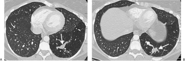 |
FIGURE 16-2. Intralobar sequestration. A: CT of a 37-year-old woman with chest pain shows a prominent tubular structure in the left lower lobe (arrow). The surrounding lung, also part of the sequestration, is hyperlucent. B: CT at a more inferior level shows the tubular structure leading to a lobulated mass (arrow) in the left lower lobe. C: CT with mediastinal windowing shows the tubular structure be a vessel (arrow). D: CT of the upper abdomen shows a prominent vessel (arrow) arising from the abdominal aorta and heading toward the left lower lobe. E: Coronal reformatted CT confirms that a vessel arises from the abdominal aorta (arrow) and heads superiorly toward the left lower lobe. F: Paddlewheel reformatted CT shows drainage from the left lower lobe mass to the left inferior pulmonary vein (arrow). |
P.266
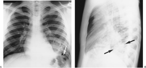 |
FIGURE 16-3. Intralobar sequestration. A:
Posteroanterior (PA) chest radiograph of a 20-year-old man with
recurrent left lower lobe pneumonia shows abnormal opacification of the
left lower lobe, with obliteration of the left hemidiaphragm shadow,
and an air–fluid level (arrow). B: Lateral view shows left lower lobe opacification involving the posterior segment (arrows). C: CT shows a slightly lobulated cystic mass in the posterior segment of the left lower lobe (arrows). D: Coronal magnetic resonance imaging (MRI) shows two arteries arising from the descending aorta (arrowheads), feeding the sequestration. E: Axial MRI shows two high-signal draining veins (arrowheads) within the sequestration (curved arrows), draining into the left inferior pulmonary vein (straight arrows). |
P.267
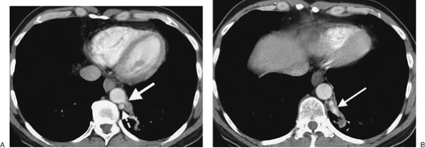 |
FIGURE 16-4. Extralobar sequestration. A:
CT of a 56-year-old man with persistent abnormal opacity in the left
lower lobe on chest radiography shows a vascular structure arising from
the descending aorta (solid arrow) and directed toward a mass in the left lower lobe. The hemiazygos vein is prominent (dashed arrow). B: CT at a more inferior level shows a large vein arising from the left lower lobe mass and draining into the hemiazygos vein (arrow). C: Coronal reformatted CT shows a prominent hemiazygos vein (arrow). D: CT at the level of the left atrium shows the hemiazygos vein (solid arrow) crossing the midline posterior to the descending aorta to join the azygos vein (dashed arrow). Surgical resection of the left lower lobe sequestration confirmed the arterial supply and venous drainage. |
P.268
Pulmonary Venolobar Syndrome
Also referred to as the scimitar syndrome or hypogenetic
lung syndrome, pulmonary venolobar syndrome is a form of partial
anomalous pulmonary venous return that is accompanied by ipsilateral
lung hypoplasia. The anomalous venous return is commonly to the
inferior vena cava (Fig. 16-5). The hypoplastic
lung (which is almost always right sided) is supplied partly or
completely by systemic arteries. The ipsilateral pulmonary artery is
diminutive. Associated cardiovascular anomalies are frequent, the most
common being atrial septal defect (6). Other
associated anomalies include pulmonary sequestration, absence of the
inferior vena cava, and accessory diaphragm. Less commonly, the
syndrome may involve tracheal trifurcation, eventration and partial
absence of the diaphragm, phrenic cyst, horseshoe lung, anomalous
superior vena cava, and absence of the left pericardium (7).
Bronchial anomalies are common, particularly isomerism (identical right
and left branching patterns). The anomalous vein is usually visible on
frontal chest radiographs as a broad, gently curved shadow descending
to the diaphragm just to the right of the heart (Figs. 16-6 and 16-7). The shadow is shaped like a Turkish sword (a scimitar); thus, the designation scimitar syndrome.
Other radiographic findings include a small ipsilateral hemithorax with
diminished pulmonary vascularity, shift of the mediastinum toward the
involved side, and, often, indistinctness of the cardiomediastinal
border on the involved side. The lateral radiograph usually
shows a retrosternal band of opacification, which is secondary to the shortening of the anteroposterior diameter of the involved lung, and contact of the anterior involved lung with a rotated and shifted mediastinum (8). Anomalous pulmonary venous return can also be an isolated finding, unassociated with other anomalies (Fig. 16-8).
P.269
shows a retrosternal band of opacification, which is secondary to the shortening of the anteroposterior diameter of the involved lung, and contact of the anterior involved lung with a rotated and shifted mediastinum (8). Anomalous pulmonary venous return can also be an isolated finding, unassociated with other anomalies (Fig. 16-8).
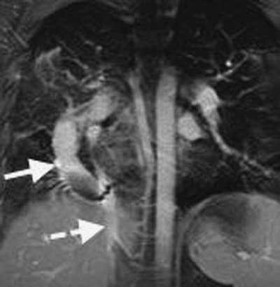 |
FIGURE 16-5. Pulmonary venolobar syndrome. MRI of a 24-year-old man shows a large venous structure (solid arrow) draining into the abdominal inferior vena cava (dashed arrow). |
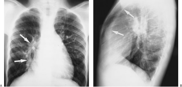 |
FIGURE 16-6. Pulmonary venolobar syndrome. A: PA chest radiograph of an 11-year-old girl shows a curvilinear band of opacification (arrows)
adjacent to the right heart border, representing an anomalous pulmonary
vein draining into the inferior vena cava. The vein is shaped like a
Turkish sword, giving rise to the name "scimitar syndrome," another
term used to describe this entity. Hypoplasia of the right lung is not
clearly seen on this view. B: Lateral view shows a retrosternal band of opacification (arrows),
created by shortening of the anteroposterior diameter of the right
lung, and contact of the anterior right lung with a rotated and shifted
mediastinum. |
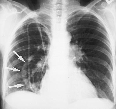 |
FIGURE 16-7. Pulmonary venolobar syndrome. PA chest radiograph of a 56-year-old woman shows the anomalous draining vein (scimitar; arrows), diminutive right pulmonary artery, and relatively small right lung. |
Pulmonary Arteriovenous Malformation
A pulmonary AVM or congenital arteriovenous fistula is
an abnormal vascular communication between a pulmonary artery and a
pulmonary vein. The etiology is thought to be defective development of
the terminal capillary loops, resulting in the formation of
thin-walled, dilated vascular spaces, usually supplied by one distended
artery and drained by one distended vein. Pulmonary AVMs are multiple
in 33% to 50% of patients and are bilateral in 8% to 20% (1).
Approximately 60% of pulmonary AVMs occur in patients with
Rendu-Osler-Weber disease (also known as hereditary hemorrhagic
telangiectasia; see also Chapter 7). Chest
radiographs and CT scans show round or oval, well-defined nodules,
which can be lobulated, ranging in size from less than 1 cm to several
centimeters in diameter, with prominent feeding and draining vessels
(see Figs. 7-32 and 7-33).
Although typically incidental findings in adults, AVMs can cause
physiologic right-to-left shunting if large, which can result in
paradoxical septic emboli.
Bronchial Atresia
Bronchial atresia is an uncommon focal obliteration of
the proximal portion of a segmental bronchus. It occurs most commonly
in the left upper lobe, followed by the left lower lobe and the right
middle lobe (9). It is thought to be related to
a vascular insult early in development. Mucoid impaction in the airway
distal to the obliterated portion is usually seen radiologically as an
ovoid, round, or branching tubular structure. The distal lung, aerated
by collateral air drift, is typically hyperlucent and hyperinflated and
has decreased vascular markings. Bronchial atresia is often discovered
incidentally. Bronchoscopy
is usually required to exclude an endobronchial neoplasm.
P.270
is usually required to exclude an endobronchial neoplasm.
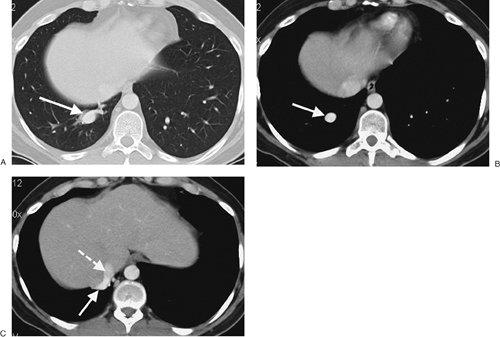 |
FIGURE 16-8. Anomalous pulmonary venous return. A: CT shows a large tubular structure in the right lower lobe (arrow). B: CT with mediastinal windowing shows enhancement of the structure (arrow), confirming its vascular nature. C: CT at a level inferior to (B) shows the vascular structure (solid arrow) draining into the inferior vena cava (dashed arrow). |
References
1. Panicek DM, Heitzman ER, Randall PA, et al. The continuum of pulmonary development anomalies. Radiographics. 1987;7:747–772.
2. Avitabile AM, Greco MA, Hulnick DH, et al. Congenital cystic adenomatoid malformation of the lung in adults. Am J Surg Pathol. 1984;8:193–202.
3. Patz EF Jr, Müller NL, Swensen SJ, et al. Congenital cystic adenomatoid malformation in adults: CT findings. J Comput Assist Tomogr. 1995;19:361–364.
4. Savic B, Birtel FJ, Tholen W, et al. Lung sequestration: report of seven cases and review of 540 published cases. Thorax. 1979;34:96–101.
5. Mata JM, Caceres J, Lucaya J, et al. CT of congenital malformations of the lung. Radiographics. 1990;10:651–674.
6. Kiely
B, Filler J, Stone S, et al. Syndrome of anomalous venous drainage of
the right lung to the inferior vena cava. A review of 67 reported cases
and three new cases in children. Am J Cardiol. 1967;20:102–115.
7. Woodring JH, Howard TA, Kanga JF. Congenital pulmonary venolobar syndrome revisited. Radiographics. 1994;14:349–369.
8. Ang JGP, Proto V. CT demonstration of congenital pulmonary venolobar syndrome. J Comput Assist Tomogr. 1984;8:753–757.
9. Kinsella D, Sissons G, Williams MP. The radiological imaging of bronchial atresia. Br J Radiol. 1992;65:681–685.